Dennis Parker, Jr, PharmD
- Neurocritical Care Clinical Pharmacist, Detroit Receiving Hospital
- Clinical Associate Professor, Eugene Applebaum College of Pharmacy and Health Sciences, Detroit, Michigan

https://cphs.wayne.edu/profile/ah2262
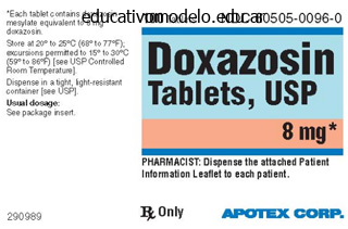
Doxazosin dosages: 4 mg, 2 mg, 1 mg
Doxazosin packs: 30 pills, 60 pills, 90 pills, 120 pills, 180 pills, 270 pills, 360 pills
Order doxazosin 1mg on-line
Tibial window chopping block assembled to the tibia template and placed flush against initial tibial ready floor and flush with the anterior tibial cortex. After drilling proximal hole and putting a stabilizing publish, the oscillating noticed is used to cut the anterior tibial window. Mark the suitable depth on the saw blade corresponding to the size of tibial element to be implanted. The appropriate mark on its upper surface have to be positioned against the anterior tibial cortex. With the correct-thickness talar pin jig in place, maintain the foot 90 degrees to the decrease leg, and insert the primary 2. The foot must be held at ninety levels relative to the lower leg; this is important during talar drill pin insertion. Align the foot into the neutral place and guide the finding runners on the talar center guide into the grooves in the tibial template superiorly and the groove in the talar flat chopping block inferiorly. Once the correct spacing is achieved, advance the talar center guide until the superior runners contact the end of the tibial template grooves. Use a center information packing between the talar heart guide and the tibial template if the house between the tibia and the talus is extreme. Advance the stop block until it meets the front of the talar flat slicing block and lock it into place using the locking screw. The two bands marked on the talar center guide correspond to two ranges throughout the six out there talar component sizes. At this point a choice have to be made whether or not the optimum talar size falls in the measurement vary 1�4 or 5�6. The anterior and posterior chamfer cuts are the identical within these respective ranges but different as the scale transitions from dimension 4 to 5. The measurement of the bearing insert part should match the size of the talar element. The measurement of the talar component and bearing insert should be smaller than or equal to that of the tibial component chosen to forestall overhang of the bearing compared to the tibial plate.
Diseases
- Crisponi syndrome
- Epilepsy microcephaly skeletal dysplasia
- Influenza
- Long QT syndrome type 2
- Bone dysplasia lethal Holmgren type
- Facioscapulohumeral muscular dystrophy
Proven doxazosin 2 mg
The reporting doctor might hesitate due to an absence of definitive information, potential accusations of interfering in the doctor-patient relationship of another, fear that skilled interactions such as affected person referrals and efficiency evaluations could also be affected, and concern of libel fits. When a doctor observes a medical error committed by another doctor, the choices include nondisclosure, recommendations to the involved doctor to disclose the error, disclosure of the error to a 3rd party corresponding to a risk-management group, or direct disclosure to the affected person. Although no strict authorized guidelines are in place, moral ideas favor actions that lead the patient to have a full understanding of what has occurred through the course of his or her medical care. Apology (as opposed to disclosure) stays a controversial side of communication following medical errors, largely because of a well-founded concern, no much less than in the past, that it could probably be used as an admission of negligence in subsequent litigation. Yet apology seems in many instances to decrease the danger of subsequent litigation, and lack of apology is a generally cited reason amongst malpractice plaintiffs for their authorized action. Usually, the partner or legally recognized home partner is considered the first-line surrogate choice maker. Commonly, the surrogate hierarchy after the spouse is kids, if all are in settlement, then mother and father, if both are in agreement, and then siblings, if all are in agreement. Incompetent sufferers may be emotionally and financially burdensome, and determination makers could have conflicts of curiosity that distort their beliefs and testament about what the patient would have wished. Studies show that sufferers and their proxies only sometimes talk about points and values involving life-sustaining applied sciences. Medical Decisions That Cannot Be Made by a Surrogate Decision Maker Some medical remedies have intense cultural connotations, might contain limitation on private freedoms such as replica, or may have historically been topic to abuse. Examples of such therapies in lots of states embrace sterilization and electroshock therapy. Many patients who categorical reluctance about resuscitation throughout surgery are frightened of burdensome outcomes, such as permanent neurologic impairment. Surgery depends on the cooperation of many caregivers with differing experience, each of whom has unbiased ethical obligations to the patient. Therefore, resuscitation agreements should be discussed with other members of the working room group. This communication prevents essential disagreements from occurring throughout a critical occasion when therapy decisions should be made quickly. Ethical distinctions between acts of omission ("letting die") and acts of commission ("killing") were and stay confusing at greatest.
Discount 2 mg doxazosin visa
Blade plate fixation of the tibiotalocalcaneal joint has been shown in biomechanical studies to have larger initial and last stiffness. However, arthritis because of malalignment, trauma, and avascular necrosis of the talus can progress relatively rapidly. The surgeon should watch the affected person strolling each toward and away from him or her and will clinically decide whether or not gait is normal or antalgic on either side. Normal ankle motion is about 50 degrees of plantarflexion and 10 to 20 degrees of dorsiflexion. Tibiotalar movement is often significantly decreased in comparison with the unaffected facet. Normal subtalar movement is about 10 to 20 degrees of inversion and 5 to 10 degrees of eversion. Subtalar movement is usually significantly decreased in comparison with the unaffected side. Past medical history may be significant for antecedent ankle or hindfoot trauma, talar osteonecrosis, diabetes, neuroarthropathy, osteochondral defect, or recurrent ankle instability. Past surgical historical past could embrace previous ankle or hindfoot surgical procedure, including open reduction and internal fixation, whole ankle arthroplasty, and former arthrodesis. Selective anesthetic injections into the ankle or subtalar joints might help to determine which joints are symptomatic. The body of the talus is saddle-shaped dorsally and fits congruently throughout the mortise created by the distal tibia and fibula. In addition, the talus and the tibial plafond are narrower posteriorly to accommodate rotation with ankle dorsiflexion and plantarflexion. The subtalar joint includes the talus and the calcaneus as they articulate via anterior, center, and posterior aspects. The major blood provide of the talar body enters retrograde via the neck of the talus, which makes the physique susceptible to avascular necrosis within the case of displaced talar neck fractures. The lateral facet of the foot is innervated by the superficial peroneal and sural nerves.
Buy doxazosin 4 mg without prescription
Alternatively: positioning within the lateral decubitus place, using a stack of folded sheets to serve as a rest for the operated leg. The duration of remedy varies based mostly on power deficiencies and the intensity of the program. Orthotic devices and shoe put on modification can be used when foot or ankle malalignment contributes to the instability. A relative contraindication for this anatomic repair is generalized ligamentous laxity as might be encountered in Ehlers-Danlos syndrome. If an osteochondral lesion is current, the ligamentous reconstruction ought to be accomplished in conjunction with arthroscopic or open treatment of the osteochondral defect. This method facilitates entry to the peroneal tendons should there be associated peroneal tendon pathology. The affected person is positioned as described, a thigh tourniquet is positioned, and a regular orthopaedic prep and drape is carried out. With the bump positioned proximal to the ankle, a dissection is carried out to isolate the inferior extensor retinaculum. The joint capsule is then incised consistent with the skin incision and simply distal to the main edge of the fibula. This inspection, along with the preoperative analysis, is used to resolve whether or not or not a restore of this ligament is required. A subperiosteal dissection is carried out on the anterior and lateral aspect of the fibula, elevating a flap three to 6 mm broad. Using curettes and rongeurs, a trough is made within the anterior and lateral aspect of the fibula at its forefront, about three mm deep and 3 mm extensive. If additional shortening is required, the capsule may be trimmed from the distal reduce edge.
Chymotrypsinum (Chymotrypsin). Doxazosin.
- Fluid retention and swelling (inflammation) associated with a broken hand and burns.
- Are there safety concerns?
- What other names is Chymotrypsin known by?
- How does Chymotrypsin work?
- Cataract surgery, when used by a healthcare professional.
- Dosing considerations for Chymotrypsin.
Source: http://www.rxlist.com/script/main/art.asp?articlekey=96417
Buy doxazosin 2mg mastercard
The incision is then carried via the subcutaneous tissues and platysma muscle using electrocautery. The superior-anterior flap is elevated to the inferior border of the parotid gland. A transverse incision is extended from the mastoid tip and is carried along the inferior border of the mandible, turning caudally and continuing alongside the anterior border of the sternocleidomastoid muscle. The incision is carried by way of the subcutaneous tissues and platysma muscle utilizing electrocautery according to the incision. Subplatysmal flaps are developed with blunt dissection methods to allow adequate mobilization of tissue. The larger auricular nerve is recognized and mobilized from the subcutaneous tissues to allow enough retraction. This will end in a small space of insensate skin but otherwise has no practical significance. For extra exposure the sternocleidomastoid can be taken down from the mastoid prominence by sectioning via the tendinous insertion. It is sometimes essential to sacrifice the higher auricular nerve; this can leave the patient with a small insensate patch of skin however no long-term useful deficit. Be sure to depart sufficient tissue cuff to allow reapproximation of the muscle on closure. C1 transverse process Internal jugular vein Middle scalene muscle Carotid sheath Deep Dissection Lymph nodes found in the area of dissection and across the spinal accent nerve can be excised. The lateral means of C1 is now simply palpable about 1 cm distal to the mastoid process. The interval between the jugular vein and the longus capitis muscular tissues is then created, permitting access to the retropharyngeal area. The retropharyngeal area could be opened additional with blunt dissection methods employing scissors, Kittners, or fingers.
Buy doxazosin 2mg otc
An oblique incision is carried out directly over the realm of the deliberate osteotomy (usually about 2 cm posterior to another concurrent incision). Fixation could be achieved via both a big axially directed screw or staples. If allograft is used, it must be ordered correctly, with sufficient size to span the space of the tendon weave (25 cm is plenty). Once the allograft is thawed, it must be bathed in antibiotic answer till prepared to be used. Consider making two separate tunnels on the posterior fibula divided by a cortical bridge between them. This will help resist the possibility of graft migration on cancellous bone within the V-shaped tunnel. Hold the foot in the desired neutral place (to about 5 degrees of overeversion), pull the graft taut, and fix it on this place. Once wounds are healed satisfactorily, the patient might begin protected weight bearing in a forged, as tolerated, for another 4 weeks. Gradual transition from cast to boot and introduction of range of movement begin 5 to 6 weeks after surgery. Rehabilitation is then instituted focusing on restoration of movement, Achilles stretching, proprioceptive coaching, and peroneal strengthening. Treatment of full rupture of the lateral ligaments of the ankle: A randomized scientific trial evaluating forged immobilization with functional therapy. Anatomic reconstruction of the lateral ankle ligaments using cut up peroneus tendon graft. Comprehensive reconstruction of the lateral ankle for chronic instability utilizing a free gracilis graft. Persistent disability with ankle sprains: A potential examination of an athletic population. Ankle stabilization with hamstring autograft: A new method using interference screws.
Cheap doxazosin 4mg on-line
Computed tomographic or magnetic resonance imaging scans are reviewed, if obtainable. Exposure of the extensor digitorum brevis muscle, sinus tarsi fats pad, and peroneal tendons. Elevation of extensor digitorum brevis and sinus tarsi fats pad as a distally based flap. Note that the objective is to preserve the normal, curved contours of the articular sides. These K-wire holes could also be additional augmented with bigger holes created via the usage of a 3-mm burr, and by feathering of the subchondral bone with a curved osteotome. Insertion of a lamina spreader and removing of the remaining medial articular cartilage. Reattachment of the extensor digitorum brevis to its origin after insertion of a tibial bone graft. This guide pin is placed fluoroscopically using axial (Harris) heel and lateral views. The preliminary information pin is often overreamed proximally (not essential with self-drilling, self-tapping screws), and a 6. Placement of the primary guide pin and screw from the apex of the calcaneal tuberosity. Placement of the second information pin and screw from the dorsomedial aspect of the talar neck. The wound is then closed using 2-0 Vicryl for the subcuticular layer and 3-0 nylon horizontal mattress sutures for the skin. The fascia overlying the anterior compartment musculature is divided according to the skin incision. After an enough quantity of cancellous graft is harvested, the window is sealed with the previously eliminated sq. plug of bone, and a layered closure of the fascia, subcutaneous tissue, and pores and skin is performed. Time from graft harvest to insertion into the fusion website must be lower than 30 minutes. Preservation of subchondral bone will present structural support and will enable for better coaptation. Countersinking of the screw heads and avoidance of a screw head positioned on the weight-bearing plantar floor of the calcaneus will reduce complaints related to the hardware.
Purchase doxazosin 2 mg on line
When performed within the manner described above, hindfoot endoscopy is a secure and reliable technique of diagnosing and treating quite a lot of posterior ankle issues. The determination to treat posterior in addition to anterior pathology is made preoperatively. After finishing the posterior process, the portals are sutured and the patient is turned and the anterior procedure is carried out. Arthroscopic excision of the os trigonum: a brand new technique with preliminary clinical results. Tenosynovitis of the flexor hallucis longus: a scientific research of the spectrum of presentation and remedy. To stop sural nerve damage you will want to create the posterolateral portal as described beforehand, near the Achilles tendon, first making a stab incision after which continuing with blunt dissection by a mosquito clamp. Avoiding the potential issues of working via a posteromedial portal, the trick is to angle the instrument (shaver, burr, punch) within the posteromedial portal at 90 levels to the arthroscope shaft. The arthroscope shaft subsequently is used as a guide for the instrument to travel into the path of the joint. In areas close to the neurovascular bundle, the aspirator must be set to a minimal quantity of suction. It could be caused by an acute or persistent injury, with the os trigonum or trigonal strategy of the talus as essentially the most offending construction. It mineralizes between the ages of eleven and thirteen years in boys and 8 and eleven years in women. It fuses with the posterior talus within 1 year, forming the posterolateral course of, usually known as the Stieda or trigonal process. In some variants, the posterior tibial artery could be skinny or absent (0�2%), with the dominant peroneal artery traversing across the posterior ankle toward the tarsal tunnel.
1mg doxazosin otc
In the transverse plane, the foot is externally rotated to the limb so that the thigh�foot axis is 10� to 15� externally rotated. In the axial plane, the calcaneal bisection line should be parallel or barely valgus (0� to 2�) and coincide with the mid-diaphyseal line of the tibia. External fixation supplies gradual correct multiplanar (rotation, angulation, and translation) realignment, while concurrently correcting limb length. Therefore, sufferers with a malunited ankle fusion, with or with no limb-length discrepancy, may be efficiently treated with minimally invasive Gigli noticed osteotomy and gradual exterior fixation correction. Treatment of malunion and nonunion on the web site of an ankle fusion with the Ilizarov apparatus. Changes seen on radiographs embody joint area narrowing, osteophytes, and subchondral bone sclerosis. The most common causes of degenerative modifications in the ankle are secondary to trauma and irregular ankle mechanics. Posttraumatic arthritis is correlated to the severity of the fracture pattern and nonanatomic reduction of articular surfaces. It is normally secondary to previous ankle fractures, talus fractures, or ligamentous instability. Rheumatoid or different inflammatory arthropathies and an infection may cause vital ankle pain, deformities, and arthritis. Options for patients who fail to respond to conservative therapy for ankle arthrosis are tibiotalar arthrodesis, complete ankle arthroplasty, and fresh ankle osteochondral shell allografts. Type three injuries also contain the subchondral bone and thus heal with a fibrocartilage, consisting mainly of type I collagen. Posttraumatic arthritis is the commonest explanation for ankle arthritis regardless of advances in open reduction and inner fixation of ankle and pilon fractures.
Real Experiences: Customer Reviews on Doxazosin
Topork, 46 years: This position also needs to be according to the tibial shaft axis in order that minimal changes shall be needed. Researchers are obligated to maximize benefits and minimize potential harms, including bodily, psychological, social, legal, and monetary harms. Microcervical foraminotomy: a surgical alternative for intractable radicular ache. Under a shared financial savings program, the provider receives a share of savings associated with decreased health care spending.
Fedor, 34 years: To permit for the distraction, the rods have to be slightly longer than will finally be needed. This spinal canal stenosis might lead to neurogenic claudication or a monoradiculopathy. The peroneal tunnel compression take a look at consists of having the patient perform this motion while palpating the posterior border of the fibula. The long arm of the inverted Y is the length that the tendon has been elongated-equal to the size of the measured gap.
Stejnar, 56 years: The mixture of recurvatum deformity above the ankle and equinus contracture of the ankle will lead to a foot translated forward place, with an extension second on the knee. Examination of the whole ankle arthroplasty for ligament instability should include the next: Medial�lateral stress radiographs. After choosing the suitable size of talar slicing block, fix it with two or three short pins. Preserved feedforward, however inhibited frontal-to-temporal connectivity, occurs in sufferers in vegetative states, however not in minimally conscious states or normal controls.
Mine-Boss, 45 years: Properly sizing the interbody implants and fully packing the disc area with graft material can help cut back the chance of this complication. If mechanical instability of the ankle is present, a ligament reconstruction on the time of the primary stage can be included. Take care not to place the osteotomy too far distal and destabilize the calcaneocuboid joint. The anterior half is eliminated, and then removal of the posterior portion could be delayed until after the posterior talar minimize is accomplished.
9 of 10 - Review by Y. Milok
Votes: 277 votes
Total customer reviews: 277
References
- Evans BK, Donley DK, Whitaker JN: Neurological manifestations of infection with the human immunodeficiency viruses. In Scheld MW, Whitley RJ, Durack DT, editors: Infections of the Central Nervous System, Raven. New York, 1991, p 201.
- Pang D, Dias MS: Ahab-Barmada M. Split cord malformation: Part I: a unified theory of embryogenesis for double spinal cord malformations, Neurosurgery 31(3):451n480, 1992.
- Huang R, Sacks J, Thai H, et al. Impact of stents and abciximab on survival from cardiogenic shock treated with percutaneous coronary intervention. Catheter Cardiovasc Interv. 2005;65:25.
- Khue PM, Truffot-Pernot C, Texier-Maugein J, et al. A 10-year prospective surveillance of Mycobacterium tuberculosis drug resistance in France 1995-2004.
- Tanaka K, Hori T, Hatakeyama N, et al. Quantification of BK polyoma viruria in Japanese children and adults with hemorrhagic cystitis complicating stem cell transplantation. J Med Virol. 2008;80(12):2108-2112.
- Grefte A, van der Giessen M, van Son W, et al. Circulating cytomegalovirus (CMV)-infected endothelial cells in patients with an active CMV infection. J Infect Dis. 1993;167:270-277.
- McCance RA, Fairweather DVI, Barrett AM, et al. Genetic, clinical, biochemical and pathological features of hypophosphatasia. Quart J Med 1956;25:523.

