Kenneth R. Lawrence, B.S., PHARM.D.
- Assistant Professor
- Department of Medicine
- Tufts University School of Medicine
- Senior Clinical Pharmacy Specialist
- Pharmacy
- Tufts Medical Center
- Boston, Massachusetts
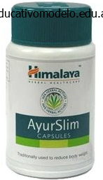
Ayurslim dosages: 60 caps
Ayurslim packs: 1 packs, 2 packs, 3 packs, 4 packs, 5 packs, 6 packs
Order ayurslim 60 caps mastercard
In a retrospective study of forty one sufferers by Cho and Sung,39 they found that affected person age was not a significant predictor of fusion failure. Importantly, the authors found that when surgical procedure was delayed for greater than 1 week, the incidence of fusion failure considerably increased with an elevated odds ratio of 37. Likewise, a fracture hole of 2 mm or extra was discovered to be considerably related to fusion failure and a 21 times great likelihood of fracture nonunion. These two components ought to definitely be considered important when considering anterior dens osteosynthesis for these fractures. In contrast, Platzer et al46 discovered some vital differences in outcomes with respect to affected person age. Six sufferers had residual neurologic deficits, of which only two had incomplete decision after conservative administration. Successful union was achieved in 96% of youthful sufferers and 88% of older sufferers. Treatment priorities ought to be individualized primarily based on affected person age, fracture pattern, associated neurologic deficits, and overall medical condition. Indications for surgical administration embody an unstable fracture pattern, high danger of nonunion, neurologic deficits, transverse ligament disruption, and contraindication to halo immobilization. There continues to be debate surrounding operative versus nonoperative strategy to odontoid fractures in addition to which surgical process is most appropriate. Posterior approaches include transarticular screw fixation, C1 lateral mass with C2 pedicle screw fixation, and posterior wiring. Studies of these strategies report high fusion rates; however, C1�C2 movement is lost. When planning the surgical strategy, careful patient selection is necessary to minimize intraoperative and postoperative problems. Site-specific issues for anterior odontoid screw fixation embrace nonunion, loss of reduction, screw pullout, airway issues, and medical sequelae from surgical procedure. Causes of mortality included cardiac arrest, respiratory failure from pneumonia, and pulmonary embolism. Other studies have also seemed at the issues of treating sufferers older than 65 years.
Generic ayurslim 60 caps buy
Often, affected patients also have an elevated threat of cancers in organs apart from the colon. Patients could have extracolonic manifestations, which include duodenal adenomas and mandibular osteomas. The adenoma�carcinoma sequence progresses much more quickly in Lynch syndrome than in sporadic colon cancer. There is an elevated danger of extracolonic malignancies, including endometrial, gastric, small bowel, renal pelvic, ureteral, and ovarian neoplasms. The screening interval is each 10 years, and screening modalities beside colonoscopy can be used. Postpolypectomy surveillance: � Advanced adenoma (see earlier) or three or extra adenomas: repeat colonoscopy in three years. Postcolorectal cancer resection surveillance: � Repeat colonoscopy 1 year after curative resection. If the examination is normal, then the interval before the following examination should be 3 years, and then 5 years thereafter if the examinations remain unfavorable for adenomas. Colorectal Neoplasms 161 Questions Questions 1 and a pair of relate to the clinical vignette firstly of this chapter. Colonoscopy revealed a 5cm mass within the ascending colon and a further 2cm mass in the descending colon. A Right hemicolectomy and endoscopic resection of the colon most cancers in the descending colon. A 74yearold man undergoes colonoscopy because of intermittent rectal bleeding and is discovered to have a 3cm pedunculated polypoid mass in the sigmoid colon. The pathology report exhibits highgrade dysplasia; no tumor cells are seen in the polyp stalk. He has no gastrointestinal complaints, and states that a flexible sigmoidoscopy accomplished roughly 5 years ago was normal.
60 caps ayurslim order free shipping
In a comprehensive systematic evaluate, Winegar et al studied the complications and outcomes related to the varied occipitocervical instrumentation methods. Screw�rod strategies offered the general greatest fusion rates; however, the underlying etiology of instability additionally appears to affect the speed of fusion. Instability at this degree might lead to ache, paresis, paralysis, respiratory misery, or death. Occipitocervical arthrodesis is a technically challenging procedure indicated in the setting of instability and in conjunction with decompressive surgery. To attain an osseous fusion, decortication of the concerned ranges with placement of bone graft and immediate inflexible internal fixation with applicable spinal instrumentation are required. Although nearly all of these procedures are efficient and safe, problems do occur. The exterior occipital protuberance, or inion, is a palpable projection superior to the apex of the nuchal line and in the midline of the squama portion of the cranium and the attachment web site of the nuchal ligament, which serves as an avascular aircraft of dissection. The inion corresponds to the intracranial torcula, which is the confluence of the transverse sinuses. The transverse sinuses correspond to the superior nuchal line, a palpable projection operating at a 43-degree angle from the horizontal cranium bottom line. The intercondylar distance decreases from anterior to posterior, with corresponding common distances of 41. The posterior side of the anterior arch of C1 articulates with the odontoid through a synovial joint. Some individuals possess an ossified posterior atlanto-occipital membrane also called an arcuate foramen or ponticulus posticus, which primarily envelopes the artery throughout the posterolateral facet of C1. This has been simulated with surgical odontoidectomies, which will increase flexion by 70. The shoulders are taped to enhance intraoperative radiographic visualization and facilitate dissection by way of posterior neck folds. Placing the affected person in a reverse Trendelenburg position facilitates venous drainage and mitigates facial swelling.
Cheap ayurslim 60 caps buy line
Lacrimal canaliculi are two curved canals that start as a lacrimal punctum (or pore) in the margin of the eyelid and open Lacrimal sac is the upper dilated end of the nasolacrimal duct, which opens into the inferior meatus of the nasal cavity. Tears enter the lacrimal canaliculi by way of their lacrimal puncta (which is on the summit of the lacrimal papilla) before the nasolacrimal duct opens into the inferior meatus is partially coated by a mucosal fold (valve of Hasner). Excess tears flow through nasolacrimal duct which drains into the inferior nasal meatus. Junction of lacrimal sac and canaliculus � Valve of Rosenmuller is a fold of mucous membrane on the junction between canaliculus and lacrimal sac. Arterial Supply (Eyeball) Ophthalmic artery is a branch of the inner carotid artery (cerebral part), enters the orbit via the optic canal beneath the optic nerve. It provides numerous ocular and orbital vessels Central artery of the retina travels in the optic nerve, divides into superior and inferior branches to the optic disk, and each of those further divides into temporal and nasal branches. Long posterior ciliary arteries (branches of ophthalmic artery) pierce the posterior a half of the sclera at some distance from the optic nerve, and run forward, between the sclera and choroid, to the ciliary muscle, the place they divide into two branches. They type an arterial circle, the circulus arteriosus main (around the circumference of the iris), from which quite a few converging branches run, within the substance of the iris, to its pupillary margin, the place they kind a second (incomplete) arterial circle, the circulus arteriosus minor. It is related to the fibrous extension of the ocular tendons (annulus of Zinn). Arteries of orbit Artery Ophthalmic Central artery of retina Course and distribution Traverses optic foramen to reach orbital cavity Pierces dural sheath of optic nerve and runs to eyeball; branches from center of optic disc; provides optic retina (except cones and rods) Supraorbital Passes superiorly and posteriorly from supraorbital foramen to supply brow and scalp Supratrochlear Passes from supraorbital margin to brow and scalp. Lacrimal Passes alongside superior border of lateral rectus muscle to supply lacrimal gland, conjunctiva, and eyelids Dorsal nasal Ophthalmic artery Courses alongside dorsal side of nostril and provides its surface Short posterior ciliaries Pierce sclera at periphery of optic nerve to supply choroid, which in flip provides cones and rods of optic retina Long posterior ciliaries Pierce sclera to provide ciliary physique and iris Posterior ethmoidal Passes via posterior ethmoidal foramen to posterior ethmoidal cells Anterior ethmoidal Passes through anterior ethmoidal foramen to anterior cranial fossa; supplies anterior and center ethmoidal cells, frontal sinus, nasal cavity, and pores and skin on dorsum of nose. Superficial temporal artery � � � � � Ophthalmic artery provides central artery of retina. It also provides the supraorbital & supratrochlear arteries, together with dorsal nasal artery. Ophthalmic artery arises from inside carotid artery because it emerges from the roof of the cavernous sinus, enters the orbit through optic canal inferolateral to the optic nerve, both mendacity in a typical dural sheath. Gives central artery to retina (an finish artery), and in addition provides ethmoidal sinuses by giving ethmoidal arteries. Leaves orbit by way of inferior orbital fissure Venous Drainage (Eyeball) Ophthalmic Veins (dig): Superior ophthalmic vein is formed by the union of the supraorbital, supratrochlear, and angular veins.
Best ayurslim 60 caps
Nerve compression causes atrophy and weak point of the muscle tissue of the hand and, in advanced circumstances, of the forearm, with ache and sensory disturbances within the arm. Thymus Thymus is a bilobed construction, mendacity within the neck anterior to the trachea and the anterior a part of the superior mediastinum (may lengthen into anterior mediastinum), attains its biggest relative measurement within the neonate, continues to develop till puberty, and then undergoes a gradual involution (replaced by fat). It is supplied by the inferior thyroid and internal thoracic artery, and produces a hormone, thymosin, which promotes T-lymphocyte differentiation and maturation. Bones are derived from somatopleuric layer of lateral plate mesoderm and muscular tissues get their origin from para-axial At weeks 7�9, the first ossification centers are seen in the clavicle, humerus, radius, and ulnar bones. Upper limbs rotate laterally by 90 degrees, in order that the thumb turns into lateral and little finger medial. The flexor compartment comes anterior and the extensor compartment becomes posterior. Ulna bone is postaxial bone with the preaxial vein becomes the cephalic vein and drains into the axillary vein in the axilla. The postaxial vein turns into the Subclavian artery represents the lateral branch of the seventh intersegmental artery. Its major continuation, the axial the original axial vessel ultimately persists as the anterior interosseous artery and the deep palmar arch. Somatic lateral plate mesoderm � Upper and lower limb bones (appendicular skeleton) develop from the somatic portion of lateral plate mesoderm, whereas muscle tissue develop from paraaxial mesoderm. A nutrient foramen is found in the lateral finish of the subclavian groove, running in a lateral path; the nutrient artery is derived from the suprascapular artery. Clavicle is the primary bone to begin ossification (between the fifth and 6th week of intrauterine life) and is the final bone to complete it (at 25 years). It ossifies largely in membrane except sternal and acromial zones (true cartilage). Most frequent site of fracture is the junction of medial 1/3rd with lateral 2/3rd � the fracture clavicle is most frequently within the center third (at the junction of lateral 1/3rd and medial 2/3rd) and leads to upward displacement of the proximal fragment pulled by the sternocleidomastoid muscle and downward displacement of the distal fragment by the deltoid muscle and gravity. Coracoid Process supplies the origin of the coracobrachialis and quick head of biceps brachii, the insertion of the pectoralis minor, and the attachment site for varied ligaments. Scapular Notch is bridged by the superior transverse scapular ligament and converted right into a foramen that transmits the suprascapular nerve.
Ayurslim 60 caps buy low price
Pulmonary veins develop in the dorsal mesocardium and open into the posterior wall of left atrium. The proper venous valve is significantly stretched out and turns into subdivided into three elements by formation of two muscular bands: the superior and inferior limbic bands. Opening of coronary sinus has an incomplete semicircular valve called the Thebesian valve, which develops from the decrease a part of the right venous valve. Septum formation in the coronary heart in part arises from development of endocardial cushion tissue in that atrioventricular canal (atrioventricular cushions) and in the cono-truncal area (cono-truncal swellings). Cushion tissue then turns into fibrous and forms the mitral (bicuspid) valve on the left and the tricuspid valve on the right. Development of Atrium and Interatrial Septum Atrial Development Atrial growth depends upon growth of the unique atrial region and incorporation of further structures. On the proper, the sinus venosus is included and varieties the smooth-walled portion of the best atrium, which is separated from the trabeculated portion by the crista terminalis. On the left, the pulmonary vein, which forms within the dorsal mesocardium, is positioned into the posterior wall of the left atrium when cells in the dorsal mesenchyme proliferate and accompany the septum primum as this structure grows toward the ground of the atrium. Development of the pulmonary vein begins in the midline after which shifts to the left. At birth, when strain in the left atrium will increase, the 2 septa press against one another and close the communication between the 2 atria, completed at round 3 months after birth. As the septum primum and septum secundum get fused with one another, foramen ovale in septum secundum is apposed and closed by septum primum. The arrows in 6 indicate the path of blood circulate from the best atrium to the left atrium throughout the fully developed atrial septum. Fossa ovalis is an oval depression on the interatrial portion consisting of the valve of the fossa ovalis (a central sheet of thin fibrous tissue) which is a remnant of septum primum. Limbus fossa ovalis is the outstanding horseshoe-shaped margin of the fossa ovalis; it represents the sting of the fetal septum secundum.
Ayurslim 60 caps order with visa
Each opens into the vestibule by a 2 cm duct, situated in the groove between the hymen and the labium minora. The epithelium of the Bartholin duct is cuboidal close to the gland, however turns into transitional and eventually stratified squamous close to the opening of the duct. Perineal body � the female exterior genitalia (or vulva/pudendum) consists of a vestibule of vagina and its surrounding structures similar to mons pubis, labia majora, labia minora, clitoris, vestibular bulb and pair of larger vestibular glands. Located on the junction of anterior 1/3 and center 1/3 of labia majora � Bartholin gland is situated at the junction of center 1/3 and posterior 1/3 of labia majora. Perineum Perineum is the diamond-shaped area between the thighs, which corresponds to the outlet of the pelvis and presents It consists of perineal pouches (superficial and deep); ischiorectal fossa; pudendal canal and anal canal. Boundaries: Anterior: Pubic symphysis, arch and the arcuate ligament Anterolateral: Ischiopubic rami Lateral: Ischial tuberosities Postero-lateral: Sacrotuberous ligaments Posterior: Tip of the coccyx Floor: Skin and fascia Roof: Pelvic diaphragm and associated fascia It is divided into an anterior urogenital triangle and a posterior anal triangle by a line drawn across the floor connecting the ischial tuberosities. Pelvis Perineal Pouches Urogenital triangle accommodates the superficial and deep perineal pouches (spaces): Superficial Perineal Pouch It lies between the perineal membrane (inferior fascia of the urogenital diaphragm) and the Colles fascia (membranous layer of superficial perineal fascia). It is an open compartment, because of the reality that anteriorly, the house communicates freely with the potential house lying between the superficial fascia of the anterior abdominal wall and the anterior abdominal muscle tissue. The superficial perineal muscles are removed within the left of the diagram to present crus and bulb of the penis. The superficial perneal muscle tissue have been removed in the left half of the diagram to present bulb of the vestibule and higher vestibular gland. Deep perineal pouch is enclosed partly by the perineum, and positioned superior to the perineal membrane (inferior fascia It lies between the superior and inferior fasciae of the urogenital diaphragm. Recently the deep pouch is being described as the region between the perineal membrane and pelvic diaphragm. It accommodates muscle tissue like external urethral sphincter and deep transverse perinei, attaching to the perineal body. The ducts move by way of the perineal membrane to attain superficial perineal pouch and open into the bulbous portion of the spongy (penile) urethra. Clinical Correlations � Episiotomy is a surgical incision of the perineum (and the posterior vaginal wall) to enlarge the vaginal opening during childbirth. Perineal Fascia Perineal fascia has two components (superficial and deep) and every of those can be subdivided into superficial and deep components.
60 caps ayurslim purchase free shipping
Primary pedicle screw augmentation in osteoporotic lumbar vertebrae: biomechanical evaluation of pedicle fixation power. Improved bone-screw interface with hydroxyapatite coating: an in vivo examine of loaded pedicle screws in sheep. Pedicular fixation in the osteoporotic spine: a pilot in vivo examine on long-term ovariectomized sheep. The "medio-latero-superior trajectory method": an alternate cortical trajectory for pedicle fixation. A new inner fixation device for disorders of the lumbar and thoracolumbar backbone. Pedicle screw insertion angle and pullout power: comparison of 2 proposed methods. Even though cortical screws are nonetheless subject to various hazards, adherence to meticulous surgical method and the correct use of diagnostic tools together with intraoperative imaging modalities and neuromonitoring can be expected to reduce the risk of serious antagonistic occasions. Cortical screws utilize a unique caudal�cephalad and medial� lateral trajectory which allows them to have interaction extra cortical bone and safe larger buy inside the vertebra, which has been confirmed by biomechanical testing. Because the instruments and implants are directed away from the thecal sac and nerve roots, the safety profile of cortical screws could also be improved relative to that of transpedicular instrumentation. Screws present inflexible constructs with the benefit of early mobilization, quick therapeutic, and high fusion rates, while obviating the necessity for exterior bracing during postoperative restoration. Therefore, quite a few decisions are available which might significantly differ when it comes to biomechanical energy, threat of damage to osseous or gentle tissue anatomic parts and long-term complication profile. Use of screw-based fixation methods, like another surgical instrumentation technique, is related to issues, and backbone surgeons should have enough knowledge about the nature of those issues, how to forestall them, and tips on how to deal with and handle them should they happen. Intraoperative complications can occur during surgical dissection or on account of inappropriate hardware positioning. Late problems include loosening, pullout, breakage of the screw or rod/plate, lack of reduction, pseudarthrosis, and adjoining section disease. This article consists of a evaluate of the obtainable literature with main concentrate on the potential problems of various sorts of screw-based fixation strategies carried out through posterior method to the backbone. Complications of Posterior Screw Fixation in Spine Surgery postsurgical angiography should be carried out. Although the unique strategy of Goel and Laheri for screw�plate constructs required sectioning of C2 nerve root,16 within the modified approach (screw�rod construct) sectioning the C2 nerve is optionally available.
Real Experiences: Customer Reviews on Ayurslim
Ressel, 27 years: Furthermore, applicable screw positioning usually poses an actual surgical problem due to its limited positioning choices. Posterior: Fascia overlaying interos- � Distally: Continuous with sei and medial three metacarpals. The agger nasi air cell is essentially the most anterior ethmoidal air cell and lies anterolateral to the frontal recess. In the radial groove, it offers five branches: Lower lateral cutaneous nerve of the arm (innervation on the lateral surface of the arm up to the elbow); posterior cutaneous nerve of the forearm (innervation down the center of the back of the forearm up to the wrist); nerve to lateral head of triceps; nerve to medial head of triceps and nerve to anconeus (passes via medial head of triceps to attain the anconeus).
Aila, 21 years: L4,L5; S1,S2,S3 � Sciatic nerve arises from the ventral divisions of L-4,5 and S-1,2,3. Microneurosurgery: Volume 1: Microsurgical Anatomy of the Basal Cisterns of the Brain, Diagnostic Studies, General Operative Techniques and Pathological Consideration of the Intracranial Aneurysms. Risks and cost-effectiveness of sublaminar wiring in posterior fusion of cervical spine trauma. Minimally invasive extracavitary method for thoracic discectomy and interbody fusion: 1-year scientific and radiographic outcomes in thirteen patients in contrast with a cohort of conventional anterior transthoracic approaches.
Josh, 36 years: The contents (in medial to lateral order) are the median nerve, brachial artery, biceps tendon and radial nerve. Thereafter, devoted angiography of the vessel to be embolized is carried out to exclude the presence of dangerous anastomoses. In the supine position, the hepatorenal pouch (of Morison) is more dependent than the best paracolic gutter. D Oxygen tension is highest near the central vein; blood flows from the central vein to the portal tract.
Grok, 41 years: Radiation is another adjunct in circumstances where complete resection is deemed too dangerous, but has been reported to increase the danger of malignant transformation. Free air is typically seen between the liver and the stomach wall on a lateral decubitus film. Stretch-induced nerve injury as a cause of paralysis secondary to the anterior cervical strategy. Long-term tumor control of benign intracranial meningiomas after radiosurgery in a sequence of 4565 patients.
Goose, 64 years: The superior thyroid arteries anastomose alongside its upper border and the inferior thyroid veins depart the thyroid gland at its lower border. Physical examination reveals an unresponsive young lady with conjunctival icterus. The radiologic study of alternative within the diagnosis of achalasia is a barium swallow (barium esophagogram) carried out beneath fluoroscopic steerage. While the superficial plexus is discovered extending from the subcutaneous layer, the deep plexus surrounds the vertebral artery and its tributaries.
Masil, 59 years: There is as well as thinning and dehiscence of parts of the bony covering of the optic (c) and carotid (a,d) canals. She is a single mother solely supporting her three children, and reviews working lengthy hours as a law associate. Hypothalamo-hypophysial portal system consists of two units of capillaries: one within the hypothalamus (median eminence) and the opposite in the hypophysis cerebri (sinusoids of pars anterior). The superior part begins at the pylorus of the abdomen (gastroduodenal junction) which is marked by the prepyloric vein.
9 of 10 - Review by L. Ballock
Votes: 335 votes
Total customer reviews: 335
References
- Chalasani N, Baluyut A, Ismail A, et al. Cholangiocarcinoma in patients with primary sclerosing cholangitis: a multicenter case-control study. Hepatology. 2000;31:7-11.
- Kramer JM, Newby LK, Chang WC, et al. International variation in the use of evidencebased medicines for acute coronary syndromes. Eur Heart J 2003;24(23):2133-2141.
- Lloyd RV, Kloppel G, Rosai J, eds. WHO Classification of Tumours of Endocrine Organs. 4th ed. Lyon, France: IARC Press; 2017.
- Granfeldt Ostgard LS, Medeiros BC, Sengelov H, et al. Epidemiology and clinical significance of secondary and therapy-related acute myeloid leukemia: a national population-based cohort study. J Clin Oncol 2015;33(31):3641- 3649.
- Gravenstein JS, Cooper JB, Orkin FK: Work and rest cycles in anesthesia practice, Anesthesiology 72:737-742, 1990.
- Kellogg ND, Menard SW, Santos A: Genital anatomy in pregnant adolescents: 'normal' does not mean 'nothing happened.' Pediatrics 113: e67-e69, 2004.
- Dunn R, Maclean A. General gastroenterology. In: Dunn R, et al, editors. The emergency medicine manual, 5th ed. Adelaide: Venom Publishing; 2010.

